Cart 0

← Early Detection

Introduction to Early Detection


The goal of screening exams for early breast cancer detection is to find cancers before they start to cause symptoms. Screening refers to tests and examinations used to detect a disease, such as cancer, in people who do not have any symptoms. Early detection means applying a strategy that results in an earlier diagnosis of breast cancer than otherwise might have occurred.

Breast cancers that are detected because they are causing symptoms tend to be relatively larger and are more likely to have spread beyond the breast. In contrast, breast cancers found during screening exams are more likely to be small and still confined to the breast.

The size of a breast cancer and how far it has spread are the most important factors in predicting the prognosis (the outlook for chances of survival) of a woman with this disease. Finding a breast cancer as early as possible greatly improves the likelihood that treatment will be successful.

There is no question that early detection tests for breast cancer save many thousands of lives each year, and that many more lives could be saved if even more women took advantage of these tests. Be Breast Aware!


Breast self-examination

Is used as a starting point for women to be aware of their breasts. However, it does not accurately determine the presence of breast cancer. Clinical breast examination is a screening method where breasts are examined by a health professional such as a doctor or nurse. Mammography screening is an effective tool for early detection.


Can you help?

Please help us with creating awareness about breast cancer and empowering and supporting people that are affected by it.


Stay in touch

Take part in our story. You can follow us on Instagram and Facebook, and Sign Up to our Email Newsletter. 


← Early Detection

Breast Change


The size and shape of women’s breasts varies considerably. Some women have a large amount of breast tissue, and therefore, have large breasts. Other women have a smaller amount of tissue with little breast fat.

A woman’s breasts are rarely balanced (symmetrical). Usually, one breast is slightly larger or smaller, higher or lower, or shaped differently than the other.

The size and characteristics of the nipple also vary greater from one woman to another. In some women, the nipples are constantly erect. In others, they will only become erect when stimulated by cold or touch. Some women also have inverted (turned in) nipples. Inverted nipples are not a cause for concern unless the condition is a new change. Since there are hair follicles around the nipple, hair on the breast is not uncommon.

The nipple can be flat, round, or cylindrical in shape. The color of the nipple is determined by the thinness and pigmentation of its skin. The nipple and areola (pigmented region surrounding the nipple) contain specialized muscle fibers that respond to stimulation to make the nipple erect. The areola also houses the Montgomery’s gland that may appear as tiny, raised bumps on the surface of the areola. The Montgomery’s gland helps lubricate the areola. When the nipple is stimulated, the muscle fibers will contract, the areola will pucker, and the nipples become hard.

Breasts change through a woman’s life. Young women have more glandular tissue than older women, making their breasts former and denser. As women grow older, the proportion of fat increases and breasts tend to droop more.

On mammogram films, breast masses, including both non-cancerous and cancerous lesions, appear as white regions. Fat appears as black regions on the films. All other components of the breast (glands, connective tissue, tumors, calcium deposits, etc.) appear as shades of white on a mammogram. In general, younger women have denser breasts. As a woman ages, her breasts become less dense and the space is filled with fatty tissue shown as dark areas on mammography x-rays. It is usually easier for radiologists to detect breast cancer in older women because abnormal areas are easier to spot.

During each menstrual cycle, breast tissue tends to swell from changes in the body’s levels of oestrogen and progesterone. The milk glands and ducts enlarge, and in turn, the breasts retain water.

During menstruation, breasts may temporarily feel swollen, painful, tender, or lumpy. Doctors recommend that women practice monthly breast self-examinations the week following menstruation when the breasts are least tender.

Factors that may influence a woman’s breast size include:

  • Volume of breast tissue
  • Family history
  • Age
  • Weight loss or gain
  • History of pregnancies and lactation
  • Thickness and elasticity of the breast skin
  • Degree of hormonal influences on the breast (particularly oestrogen and progesterone)
  • Menopause


Can you help?

Please help us with creating awareness about breast cancer and empowering and supporting people that are affected by it.


Stay in touch

Take part in our story. You can follow us on Instagram and Facebook, and Sign Up to our Email Newsletter. 


← Early Detection

Breast Self Examination (BSE)


BSE is a visual and physical examination for any changes in the breasts and underarms which you or a trusted person can perform on you.

Age 20 – 39

  • Do Breast Self-Examination (BSE) once a month, between the 7-10 day from the start of your period.
  • Do Clinical Breast Examination (CBE) once every 3 years, though some recommend that you do it once a year upon reaching 30 years old.

Age 40 and above

  • Do monthly BSE. For women who have reached menopause, do it on a specific date of the month.
  • Do CBE and mammogram once a year.


Breast changes.jpg


Being breast aware means knowing how your breasts usually look and feel. Look for changes in the:


Breast size and shape, pain and lump

  • An increase in size of one breast.
  • One breast has suddenly shifted position/height.
  • Unusual thickening of the breast skin.
  • A painless lump in the breast or armpit.
  • Unexplained pain in the breast with no lump.


Skin

  • Swelling, redness or distortion of the breast skin.
  • Puckering or dimpling in the skin of the nipples or breast (skin texture similar to an orange peel)
  • A sore or ulcer on the breast skin that does not heal.


Nipple

  • Sticky or bloody nipple discharge.
  • Scaling, crusting or erosion of the nipple.
  • Inversion or retraction of the nipple or area surrounding the nipple (areola)


Performing regular BSE is one way to increase your familiarity with your breasts. The key to BSE is to spot changes in your breasts. Look for changes that persist after your menstrual cycle or any changes that concern you.


BSE.gif

Standing Up

Stand undressed in front of a mirror. Look at your breasts and check each breast for anything unusual, such as puckering, dimpling, scales or changes in size, shape or symmetry. Remember to look beneath each breast, using your hands to lift the breasts if necessary.


Look

Look for breast changes at three positions:

  1. Visually inspect your breasts, arms at the side, while facing forward and while turning from side to side.
  2. Then, clasp your hands behind your head and press them forward. You should feel your chest muscles tightening, allowing you to see the contours of your breasts. Turn from side to side so you can see the outer surfaces.
  3. Next, inspect your breasts while pressing your hands firmly on your hips and bending forward slightly, pulling your shoulders and elbows forward as well.


Feel

Standing erect again, raise one arm, and use the pads of the three middle fingers of the opposite hand to feel the breast. You can use lotion or powder to help your fingers glide easily over the skin, or you can do the raised-arm part of the exam in the shower with soapy skin.


Lying Down

Lie down with a pillow under your left shoulder, and put your left hand behind your head.


Feel

Feel your left breast with the pads of the three middle fingers of your right hand. Start at the outer edge and work inward toward the nipple. Then squeeze your nipple gently to look for discharge. Do the same thing to your right breast with a pillow under your right shoulder. Be sure to include the area up to your collarbone and out to your armpit.

Feel the tissue by pressing your fingers in small, overlapping areas about the size of a 10sen coin. To be sure you cover your whole breast, take your time and follow a definite search pattern - wedge, circle or lines, to help you perform BSE more thoroughly. You will be treated by a team of professionals, including some or all of the following:

  • General Practitioners (GP) – The one who usually arranges your first referral to a specialist breast clinic.
  • Oncologist – A doctor who specializes in the diagnosis, treatment and rehabilitation of cancer patients. A clinical oncologist is trained in giving radiotherapy and chemotherapy, while a medical oncologist administers chemotherapy only.
  • Radiologist – A medically qualified doctor who specializes in the use of imaging techniques (X-rays, ultrasound, CT etc) for diagnosis, and the one who will interpret your mammogram.
  • Pathologist – A doctor specializing in identifying diseases by studying cells and tissues under a microscope.
  • Radiographer – A non-medically qualified person who assists the radiologist in imaging and is trained in using X-ray machines. Some also give radiotherapy.
  • Breast Surgeon – A specialist in breast surgery who also does fine needle aspiration and biopsy. Some may also do reconstructive surgery.
  • Breast-care nurse – Nurse with special training in dealing with breast disease, who offers information, support and advice.
  • Ward nurses – A nurse who plans your care on the ward.
  • Physiotherapist – Gives you exercises to do after surgery for you to regain your strength and fitness.
  • Psychologist – Professional counselling that can help you cope with your illness, and deal with decisions about treatment, depression, anxiety as well as family issues.
  • Social worker – Can help you work out practical difficulties including transport, childcare and financial issues


Watch the Breast Self Examination Video


Can you help?

Please help us with creating awareness about breast cancer and empowering and supporting people that are affected by it.


Stay in touch

Take part in our story. You can follow us on Instagram and Facebook, and Sign Up to our Email Newsletter. 


← Early Detection

BSE Search Pattern


To use the "wedge" pattern, start at the outer edge of the breast, and move your fingers toward the nipple and back to the edge. Check the entire breast by covering one small wedge at a time.

You can also use a "circle" pattern by beginning at the outer edge of your breast and moving your fingers slowly around the whole breast in a circle. Keep feeling the breast in smaller circles, gradually working toward the nipple.

To use the "line" technique, start under the arm and move your fingers downward slowly until they are below the breast. Then move your fingers slightly toward the middle, and slowly move back up. Continue going up and down until you cover the whole breast.

Whichever pattern you prefer, remember to apply it up to the collar bone and out to the armpit.


FAQ


What do you do if you find a lump?

One of the most important reasons to perform regular BSE is so that you know how your breasts look and feel under normal circumstances.

If you discover a lump in one breast or feel something "different" in the tissue, examine the same spot in the opposite breast. Usually, if the same area in the opposite breast feels the same there is little need for worry. However, if they feel different, or you feel a definite lump, there may be valid reason for concern and it is important to contact your doctor right away. Sometimes, the lumpiness may be due to menstrual changes, however, if you have nipple discharge or skin changes such as dimpling or puckering, your doctor may want to see you right away.

It is natural to be frightened when discovering a lump, but do not let the prospect of cancer delay you from taking action. Remember that 80 percent of all breast lumps are benign (non-cancerous).


Is BSE really an effective way to detect cancer?

Women who perform regular BSE find 90% of all breast lumps. Good technique is crucial: research shows that women who perform BSE correctly (inspecting the breasts visually and palpating with the pads of their middle three fingers) are less likely to die of breast cancer or have metastasis (cancer spread) to other parts of the body. Furthermore, women who report performing BSE thoroughly are about 35% less likely to develop advanced breast cancer.

There is nothing you can do that will absolutely prevent breast cancer. However, monthly BSE and periodic mammography and check-ups by your doctor can both lower your risk and promote early detection. Combining these measures with a healthy diet and regular exercise will also reduce your risk, and increase your peace of mind.


Can you help?

Please help us with creating awareness about breast cancer and empowering and supporting people that are affected by it.


Stay in touch

Take part in our story. You can follow us on Instagram and Facebook, and Sign Up to our Email Newsletter. 


← Early Detection

Clinical Breast Examination


What?

Clinical breast examination (CBE) is when a doctor or a nurse examines the breasts and helps find lumps that women may miss with their own self-exams.

While it's true that most lumps are found by women themselves, the abnormality in a breast can be so difficult to feel that only someone with experience would recognize it. Lumps, thickening, asymmetry—changes in your breasts that you may not notice or think are "normal"—may be detected by a doctor who examines many breasts regularly. Studies show that regular self-exam, COMBINED with an annual exam by a doctor, improves the chances of detecting cancer early.


When?

Age 20 – 39
Do Breast Self-Examination (BSE) once a month, between the 7-10 day from the start of your period
Do Clinical Breast Examination (CBE) once every 3 years

Age 40 and above
Do monthly BSE. For women who have reached menopause, do it on a specific date of the month.
Do CBE and mammogram once a year


How?

CBE is done by a trained health professional, i.e. a doctor or a nurse. You will need to remove any jewelry that might interfere with the examination. You will need to take off your clothes above the waist, and you will be given a cloth or paper gown to wear during the examination.

First, the health professional will ask you questions about any symptoms you may have, your medical history, and your risk factors for breast cancer. Tell them if you:

  • Have noticed a new lump or change in your breasts.
  • Are or might be pregnant.
  • Are breast-feeding.
  • Have breast implants.
  • Have previously had a breast biopsy.
  • Have completed menopause.
  • Are taking hormone replacement therapy.
  • Have a personal or family history of breast cancer.

Your health professional will then examine each breast, underarm, and collarbone area for changes in breast size, skin changes, and signs of injury or infection, such as bruising or redness. You may be asked to lean forward and press your hands together to tighten the muscle beneath each breast during this part of the examination.

Your health professional will feel (palpate) each breast for any unusual or painful areas or a dominant lump. A dominant lump in the breast is any lump that is new, larger, harder, or different in any other way from other lumps or the rest of the breast tissue.

Your health professional will gently press on the breast tissue from about 1 in.(2.5 cm) below the breast up to the shoulder bone. He or she also will examine your armpit (axillary area) and your neck for swollen glands (lymph nodes).

A CBE normally does not cause any discomfort. If your breasts are tender because your menstrual period is about to begin, a CBE may cause slight discomfort when the health professional presses on your breasts to feel for lumps.


Findings of a CBE may include the following:

Clinical breast examination

Normal: The nipples, breast tissue, and areas around the breast are normal in size, shape, and appearance. One breast may be slightly larger than the other. A small area of firm tissue may be present in the lower curve of the breast below the nipple. Tenderness or lumpiness that occurs in both breasts and improves after a menstrual period is normal for many women. Many women have symmetrical lumpiness or thickening in both breasts throughout the menstrual cycle. A clear or milky discharge (galactorrhea) may be present when the nipple is squeezed. This may be caused by nursing, breast stimulation, hormones, or some other normal cause.

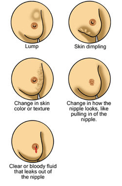
Abnormal: A firm lump or area of thickening may be present in one of your breasts.
Changes in the color or feel of your breast or nipple may be present. This can include wrinkling, dimpling, thickening, or puckering or an area that feels grainy, stringy, or thickened. A nipple may sink into the breast. A red, scaly rash or sore may be found on the nipple. Redness or warmth over a painful lump or over an entire breast may be present. This may be caused by an infection (abscess or mastitis) or cancer, such as inflammatory breast cancer. A bloody or milky discharge (galactorrhea) may occur without stimulation (spontaneous).

If a breast problem is found, the next step depends on the problem:

·   Cyclic breast pain, fibrocystic changes, or cysts may just be rechecked to see whether they change or go away on their own. Cysts may also be examined by ultrasound or drained with a needle (aspirated) to make sure they are cysts and to help relieve pain.

·   A mammogram or ultrasound may be ordered if a lump is found. Fluid may be drained if a cyst is present. Tissue for examination in the laboratory may be collected with a needle (needle aspiration or core biopsy) or through a small incision (biopsy).

·   Nipple discharge, especially if it is spontaneous or bloody, may be examined under a microscope for unusual cells.


FAQ


Are there any risks involved in doing a CBE?
There are no risks involved in doing a CBE.

Is a CBE sufficient to check on breast cancer?
A normal CBE does not guarantee that breast cancer is not present. Depending on your age and your personal and family history of breast cancer, your doctor may suggest other tests, such as a mammogram.

What affects the test of CBE?

  • The changes in your body that occur during your menstrual cycle may produce irregular changes in the structure and density of your breasts.
  • Scheduling a clinical breast examination at different times in your menstrual cycle may make it difficult to compare results of one examination with those of another.
  • If your breasts become tender just before you start your period, you may find it difficult to have a clinical breast examination at that time.
  • Fibrocystic lumps may make a clinical breast examination difficult because lumps occur throughout the breast. The fibrocystic lumps can also become tender before menstruation.


Can you help?

Please help us with creating awareness about breast cancer and empowering and supporting people that are affected by it.


Stay in touch

Take part in our story. You can follow us on Instagram and Facebook, and Sign Up to our Email Newsletter. 


← Early Detection

Positron Emission Tomography (PET)


What?

Positron emission tomography, also called PET imaging or a PET scan, is a diagnostic examination that uses signals emitted by radioactive tracers to construct images of the distribution of the tracers (positrons) in the human body. Positrons are tiny particles emitted from a radioactive substance administered to the patient. The subsequent images of the human body developed with this technique are used to evaluate a variety of diseases.


When? What are some common uses of the procedure?

PET scans are used most often to detect cancer and to examine the effects of cancer therapy by characterizing biochemical changes in the cancer. These scans can be performed on the whole body. PET scans of the heart can be used to determine blood flow to the heart muscle and help evaluate signs of coronary artery disease. PET scans of the heart can also be used to determine if areas of the heart that show decreased function are alive rather than scarred as a result of a prior heart attack, called a myocardial infarction. Combined with a myocardial perfusion study, PET scans allow differentiation of nonfunctioning heart muscle from heart muscle that would benefit from a procedure, such as angioplasty or coronary artery bypass surgery, which would reestablish adequate blood flow and improve heart function. PET scans of the brain are used to evaluate patients who have memory disorders of an undetermined cause, suspected or proven brain tumors or seizure disorders that are not responsive to medical therapy and are therefore candidates for surgery.


What does the equipment look like?

You will be taken to an examination room that houses the PET scanner, which has a hole in the middle and looks like a large doughnut. Within this machine are multiple rings of detectors that record the emission of energy from the radioactive substance in your body and permit an image of your body to be obtained. While lying on a cushioned examination table, you will be moved into the hole of the machine. The images are displayed on the monitor of a nearby computer, which is similar in appearance to the personal computer you may have in your home.


How? How does the procedure work?

Before the examination begins, a radioactive substance is produced in a machine called a cyclotron and attached, or tagged, to a natural body compound, most commonly glucose, but sometimes water or ammonia. Once this substance is administered to the patient, the radioactivity localizes in the appropriate areas of the body and is detected by the PET scanner.

Different colors or degrees of brightness on a PET image represent different levels of tissue or organ function. For example, because healthy tissue uses glucose for energy, it accumulates some of the tagged glucose, which will show up on the PET images. However, cancerous tissue, which uses more glucose than normal tissue, will accumulate more of the substance and appear brighter than normal tissue on the PET images.


How should I prepare for the procedure?

PET is usually done on an outpatient basis. Your doctor will give you detailed instructions on how to prepare for your examination. You should wear comfortable, loose-fitting clothes. You should not eat for four hours before the scan. You will be encouraged to drink water. Your doctor will instruct you regarding the use of medications before the test.

Note: Diabetic patients should ask for any specific diet guidelines to control glucose levels during the day of the test.

A nurse or technologist will take you into a special injection room, where the radioactive substance is administered as an intravenous injection (although in some cases, it will be given through an existing intravenous line or inhaled as a gas). It will then take approximately 30 to 90 minutes for the substance to travel through your body and accumulate in the tissue under study. During this time, you will be asked to rest quietly and avoid significant movement or talking, which may alter the localization of the administered substance. After that time, scanning begins. This may take 30 to 45 minutes.

Some patients, specifically those with heart disease, may undergo a stress test in which PET scans are obtained while they are at rest and again after undergoing the administration of a pharmaceutical to alter the blood flow to the heart.

Usually, there are no restrictions on daily routine after the test, although you should drink plenty of fluids to flush the radioactive substance from your body.


FAQ


What will I experience during the procedure?

The administration of the radioactive substance will feel like a slight pinprick if given by intravenous injection. You will then be made as comfortable as possible before you are positioned in the PET scanner for the test. You will be asked to remain still for the duration of the examination. Patients who are claustrophobic may feel some anxiety while positioned in the scanner. Also, some patients find it uncomfortable to hold one position for more than a few minutes. You will not feel anything related to the radioactivity of the substance in your body.


Who interprets the results and how do I get them?

Patients undergo PET because their referring physician has recommended it. A radiologist who has specialized training in PET will interpret the images and forward a report to your referring physician. It usually takes one to three days to interpret, report and deliver the results. In order to facilitate interpretation, you may be asked to bring any previous radiologic images with you, such as recent CT (CAT) scans or MRI images.


What are the benefits vs. risks?

  • Because PET allows study of body function, it can help physicians detect alterations in biochemical processes that suggest disease before changes in anatomy are apparent with other imaging tests, such as CT or MRI.
  • Because the radioactivity is very short-lived, your radiation exposure is low. The substance amount is so small that it does not affect the normal processes of the body.
  • The radioactive substance may expose radiation to the fetus in patients who are pregnant or the infants of women who are breast-feeding. The risk to the fetus or infant should be considered in relation to the potential information gain from the result of the PET examination. If you are pregnant, you should inform the PET imaging staff before the examination is performed.


What are the limitations of Positron Emission Tomography?

PET can give false results if a patient's chemical balances are not normal. Specifically, test results of diabetic patients or patients who have eaten within a few hours prior to the examination can be adversely affected because of blood sugar or blood insulin levels.

Also, because the radioactive substance decays quickly and is effective for a short period of time, it must be produced in a laboratory near the PET scanner. It is important to be on time for the appointment and to receive the radioactive substance at the scheduled time. PET must be done by a radiologist who has specialized in nuclear medicine and has substantial experience with PET. Most large medical centers now have PET services available to their patients. Medicare and insurance companies cover many of the applications of PET, and coverage continues to increase.

Finally, the value of a PET scan is enhanced when it is part of a larger diagnostic work-up. This often entails comparison of the PET scan with other imaging studies, such as CT or MRI.


Can you help?

Please help us with creating awareness about breast cancer and empowering and supporting people that are affected by it.


Stay in touch

Take part in our story. You can follow us on Instagram and Facebook, and Sign Up to our Email Newsletter.